5.Evaluation of lymphedema
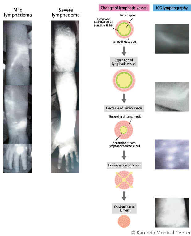
In evaluation of lymphedema, an important point of subsequent successful treatment is to know not only appearance/volume of an affected extremity and characteristic of edema, but also how function of lymphatic vessels is maintained (see the third column).
Typical test methods to evaluate function of lymphatic vessels are lymphoscintigraphy using a radioactive isotope, single photon emission computed tomography (SPECT)-CT, indocyanine green (ICG) fluorescence lymphography using contrast, and MRI lymphography. It is important to know individual conditions using a combination of these test methods, considering various condition and severity.
In our department, ICG lymphography is performed in an outpatient examination room on a usual outpatient visit to evaluate function and severity of lymphatic vessels. However, for cases in which evaluation with only ICG lymphography is insufficient, or cases in which there is a remarkable difference between the test result of ICG lymphography and symptoms, other tests are additionally performed (see "Procedure for an appointment of the outpatient service/visit/treatment of lymphedema).
Because ICG lymphography uses only 1 mL or less of contrast without radiation equipment, it is the most minimally invasive test among tests with contrast for lymphedema. This test can show clinical severity of lymphedema as well as predict changes in lymphatic vessels; therefore, it plays a very important role in considering strategies of conservative therapy or surgery.