
microbiology round
2024年5月23日のMicrobiology roundは、hypermucoviscous Klebsiella pneumoniae (hmKp)を取り上げました。
ポイントは、
・K. pneumoniaeは、hypervirulent(高病原性) K. pneumoniae(hvKp)とclassical K. pneumoniae(cKp)の2つの病原型がある。
・高齢者で基礎疾患がある患者の尿路感染、肺炎、菌血症などを起こすが、hvKpは若年の市中発症が多く、胆道疾患を伴わない肝膿瘍や、播種病変(眼内炎や髄膜炎、脳膿瘍などを含む)を起こす。
・“hypervirulent”の定義は複数の病原因子の保有していることであるが、遺伝子学的な検査は多くの施設では難しい。hypermucoviscous Klebsiella pneumoniae (hmKp)がhvKpと相関しているので、hmKpを診断するstring testを補助的に用いる。
【歴史】
・“Klebsiella”=ドイツの微生物学者の「Edwin Klebs」に敬意を表して命名された。“pneumoniae”=「pneumonia (肺炎)」
・Edwin Klebsは1875年、肺炎で死亡した患者の気道の検体中にKlebsiella pneumoniaeを初めて発見し、Carl Friedlanderが1882年にこの種を正式に記載した(1)。1887年にTrevisanによって命名された。K. pneumoniae には亜種が存在し、K. pneumoniae subsp. pneumoniae、K. pneumoniae subsp. ozaenae、K. pneumoniae subsp. rhinoscleromatisの3亜種に分けられる。DNAは相同であるが、生化学反応は異なる。
【微生物学】2)
・Klebsiella属は、LPSN(List of Prokaryotic names with Standing in Nomenclature)(2)では 27菌種、8亜種が記載されている。ヒトの感染症に主に関連するのは、K. pneumoniae、K. oxytocaとK. granulomatisである。
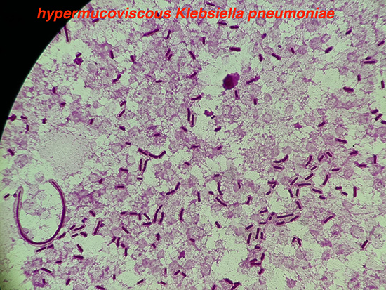
・大きさ 0.3~1.5×0.6~6 μmの通性嫌気性グラム陰性桿菌。乳糖を分解し、非運動性。粘稠性のあるコロニーを形成する。indole pyruvic acid (IPA)反応陰性、H2S陰性、インドール陰性、Voges-Proskauer(VP)反応陽性(※K. pneumoniae subsp. ozaenaeとK. pneumoniae subsp. rhinoscleromatisはVP反応陰性)。
【K.pneumoniaeの一般的な臨床像】(3)
・一般的に環境中に存在するが、ヒトの鼻咽頭や皮膚、腸管内に無症候性で定着することがある(4–6)。
・1949年に発表されたK. pneumoniaeによる市中肺炎の特徴について記述した論文では、臨床的には急速に進行する悪寒や呼吸困難、咳嗽、血液と粘液の混じった粘着性のある喀痰が出現し、胸部X線で早期の膿瘍形成や、病変のある肺葉の膨張結果として葉間裂の伸展を示す“bulging fissure sign”が特徴として記述されている (7)。
・K. pnaumoniaeの腸管への保菌はK. pneumoniaeによる感染症のリスクであり、保菌していない患者と比較して約6.9倍上昇するとの報告もある(8)。
・多くのK. pnaumoniae感染症は、ICUを中心(9)とした院内感染(10–13)であるが、健常なヒトにも感染を起こす。一般的に遭遇する感染症は尿路感染症、肺炎、菌血症であり、肝膿瘍やその他の腹腔内感染、創部感染、脳外科術後の髄膜炎などがある(5,14,15)。市中発症の髄膜炎は、肝膿瘍に伴う播種病変以外では一般的にはみられない(16,17)。
・アルコール使用、糖尿病、悪性腫瘍、末期腎不全、免疫不全などの複数の基礎疾患があることがリスク因子(5,14,15)。
【hypervirurent K. pneumoniae (hvKp)について】
・K. pneumoniaeは、hypervirulent K. pneumoniae(hvKp)とclassical K. pneumoniae(cKp)の2つの病原型に大別される。hypervirulent (高病原性)=hypermucoviscous (高粘稠性)ではなく、“hypervirulent”の定義は複数の病原因子の保有していることである(後述)。
■hvKpとcKpの臨床像の違い(18,19)
・hvKp:患者背景としては、年齢差は比較的若く、基礎疾患がないことも多い。市中発症が多く、アジア系、太平洋諸島や、ヒスパニック系での報告が多い。胆道疾患を伴わない肝膿瘍や、複数の播種病変(眼内炎や髄膜炎、脳膿瘍などを含む)を起こす。
・cKp:高齢者で、複数の基礎疾患がある患者が多い。医療曝露歴のある状況での感染症が多く、人種差はない。感染巣は単一で、播種病変をつくることは少ない。
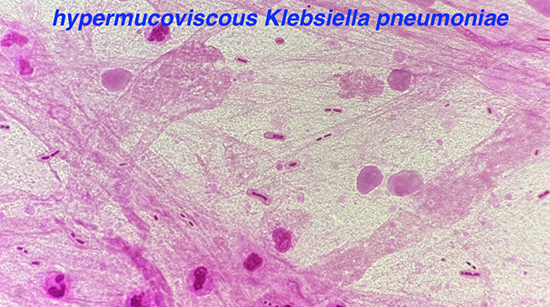
・台湾では1980年代から、特に糖尿病がある患者でK. pneumoniae単一による市中発症の肝膿瘍の症例が増加していることが認識されており、一部の症例では播種性の髄膜炎や眼内炎の合併があった(20)。その後、これらの症例の菌株の多くが、固形培地上のコロニーで高粘稠性を有する(hypermucoviscous)株であることが報告された(21)。
■hvKpの病原因子
・莢膜の血清型:
K. pneumoniaeの莢膜は、多糖体の抗原型(K血清型)に基づき77種類以上に分類され(22)、免疫細胞による貪食を回避する働きがある。台湾やシンガポールのK. pneumoniaeによる肝膿瘍の症例を対象としたコホート研究では、K1株であることが播種性感染症の合併を有するリスク因子であった(23,24)。その後、K1以外にK2が高病原性と最も関連していることが判明しており、他にK5、K16、K20、K28、K54、K57、K63も高病原性と関連づけられている(25)。
・過粘稠表現型:
コロニーの高粘稠性と関連が高いとされるrmpA(regulator of mucoid phenotupe A)遺伝子やmagA遺伝子(mucoviscosity associated gene A)が、高病原性と関連している(26)。
・Siderophores:
Klebsiella属などの腸内細菌目細菌は、自己増殖のために鉄の取り込みが必要である(27)ため、鉄を獲得するためにいくつかのsiderophores (aerobactinやsalmochelin、enterobactinやyersiniabactin)を産生する (28,29)。hvKpはcKpと比較して6~10倍のsiderophore活性を示すことが実証されている(28,30)。
■日本でのhvKp
これまでの日本国内からのK. pneumoniae菌血症の症例の報告では、遺伝学的にhvKpの定義を満たす菌株(rmpA、iuc、iroなどの遺伝子を有する菌株)は18.6〜25%である(31,32)。台湾や中国、韓国などと共にhvKpの検出頻度が高い国であることが示唆されている(31)。
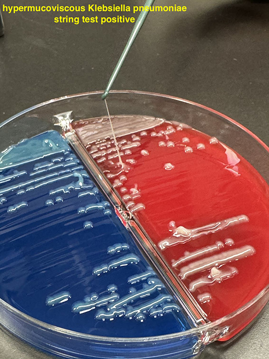
■Hypermucoviscosity (高粘稠性)と高病原性
・高粘稠性の定義は、固形培地上のコロニーを微生物検査用のループで触れて検査(string test)した時に、5mm以上の粘性の糸が形成されることと定義された(33)。string test陽性は、感度69.2%、特異度89.5%で遺伝学的な高病原性株と相関していた(31)
【治療】
・K. pneumoniaeはSHV-1などのクラスA βラクタマーゼを産生するため(34)、アンピシリンには自然耐性である。また、Extended Spectrum β-Lactamase (ESBL)やカルバペネマーゼをコードするプラスミドを保有する、最も一般的な菌の一つである(3)。
・これまでhvKPの抗菌薬感受性は比較的良好との報告が多かった(25)が、ここ数年で中国での多剤耐性のhvKpの出現(35,36)や世界的な拡散(37)が報告されている。rmpA2などの病原性遺伝子とカルバペネマーゼ遺伝子blaKPC-2の両方が組み込まれたプラスミドを持つ、hybrid plasmidも報告されている(38)。
・台湾の単施設におけるK. pneumoniae(全てセファゾリンに感受性がある株)による肝膿瘍の後方視的解析では、セファゾリンでの治療は、広域セフェム(セフトリアキソン、セフタジジムやセフォタキシム)で治療した場合と比較して、播種性感染症(特に眼内炎)や肺合併症(機械換気が必要な呼吸不全や急性呼吸窮迫症候群、敗血症性肺塞栓症)といった合併症が多かったと報告されている(39)。
1. C. Friedlaender. Ueber die Schizomyceten bei der acuten fibrösen Pneumonie. Archiv für pathologische Anatomie und Physiologie und für klinische Medicin. 1882 Feb;87:319–24.
2. LPSN - List of Prokaryotic names with Standing in Nomenclature [Internet]. [cited 2024 May 29]. Klebsiella pneumoniae. Available from: https://lpsn.dsmz.de/species/klebsiella-pneumoniae
3. Bennett JE, M. Mandell, Douglas, and Bennett’s Principles and Practice of Infectious Diseases. Ninth. Vol. 218. 2020.
4. Conlan S, Kong HH, Segre JA. Species-level analysis of DNA sequence data from the NIH Human Microbiome Project. PLoS One. 2012;7(10):e47075.
5. Siu LK, Yeh KM, Lin JC, Fung CP, Chang FY. Klebsiella pneumoniae liver abscess: a new invasive syndrome. Lancet Infect Dis. 2012 Nov;12(11):881–7.
6. Farida H, Severin JA, Gasem MH, Keuter M, van den Broek P, Hermans PWM, et al. Nasopharyngeal carriage of Klebsiella pneumoniae and other Gram-negative bacilli in pneumonia-prone age groups in Semarang, Indonesia. J Clin Microbiol. 2013 May;51(5):1614–6.
7. FELSON B, ROSENBERG LS, HAMBURGER M. Roentgen findings in acute Friedländer’s pneumonia. Radiology. 1949 Oct;53(4):559–65.
8. Gorrie CL, Mirceta M, Wick RR, Edwards DJ, Thomson NR, Strugnell RA, et al. Gastrointestinal Carriage Is a Major Reservoir of Klebsiella pneumoniae Infection in Intensive Care Patients. Clin Infect Dis. 2017 Jul 15;65(2):208–15.
9. Vincent JL, Rello J, Marshall J, Silva E, Anzueto A, Martin CD, et al. International study of the prevalence and outcomes of infection in intensive care units. JAMA. 2009 Dec 2;302(21):2323–9.
10. Weiner LM, Webb AK, Limbago B, Dudeck MA, Patel J, Kallen AJ, et al. Antimicrobial-Resistant Pathogens Associated With Healthcare-Associated Infections: Summary of Data Reported to the National Healthcare Safety Network at the Centers for Disease Control and Prevention, 2011-2014. Infect Control Hosp Epidemiol. 2016 Nov;37(11):1288–301.
11. Pitout JDD, Nordmann P, Poirel L. Carbapenemase-Producing Klebsiella pneumoniae, a Key Pathogen Set for Global Nosocomial Dominance. Antimicrob Agents Chemother. 2015 Oct;59(10):5873–84.
12. Munoz-Price LS, Poirel L, Bonomo RA, Schwaber MJ, Daikos GL, Cormican M, et al. Clinical epidemiology of the global expansion of Klebsiella pneumoniae carbapenemases. Lancet Infect Dis. 2013 Sep;13(9):785–96.
13. Zhanel GG, Adam HJ, Baxter MR, Fuller J, Nichol KA, Denisuik AJ, et al. Antimicrobial susceptibility of 22746 pathogens from Canadian hospitals: results of the CANWARD 2007-11 study. J Antimicrob Chemother. 2013 May;68 Suppl 1:i7-22.
14. Pagano L, Caira M, Trecarichi EM, Spanu T, Di Blasi R, Sica S, et al. Carbapenemase-producing Klebsiella pneumoniae and hematologic malignancies. Emerg Infect Dis. 2014 Jul;20(7):1235–6.
15. Schwaber MJ, Klarfeld-Lidji S, Navon-Venezia S, Schwartz D, Leavitt A, Carmeli Y. Predictors of carbapenem-resistant Klebsiella pneumoniae acquisition among hospitalized adults and effect of acquisition on mortality. Antimicrob Agents Chemother. 2008 Mar;52(3):1028–33.
16. Tang LM, Chen ST. Klebsiella pneumoniae meningitis: prognostic factors. Scand J Infect Dis. 1994;26(1):95–102.
17. Durand ML, Calderwood SB, Weber DJ, Miller SI, Southwick FS, Caviness VS, et al. Acute bacterial meningitis in adults. A review of 493 episodes. N Engl J Med. 1993 Jan 7;328(1):21–8.
18. Russo TA, Marr CM. Hypervirulent Klebsiella pneumoniae. Clin Microbiol Rev. 2019 Jun 19;32(3).
19. Kocsis B. Hypervirulent Klebsiella pneumoniae: An update on epidemiology, detection and antibiotic resistance. Acta Microbiol Immunol Hung. 2023 Dec 7;70(4):278–87.
20. Wang JH, Liu YC, Lee SS, Yen MY, Chen YS, Wang JH, et al. Primary liver abscess due to Klebsiella pneumoniae in Taiwan. Clin Infect Dis. 1998 Jun;26(6):1434–8.
21. Fang CT, Chuang YP, Shun CT, Chang SC, Wang JT. A novel virulence gene in Klebsiella pneumoniae strains causing primary liver abscess and septic metastatic complications. J Exp Med. 2004 Mar 1;199(5):697–705.
22. Clegg S, Murphy CN. Epidemiology and Virulence of Klebsiella pneumoniae. Microbiol Spectr. 2016 Feb;4(1).
23. Fung CP, Hu BS, Chang FY, Lee SC, Kuo BI, Ho M, et al. A 5-year study of the seroepidemiology of Klebsiella pneumoniae: high prevalence of capsular serotype K1 in Taiwan and implication for vaccine efficacy. J Infect Dis. 2000 Jun;181(6):2075–9.
24. Yeh KM, Kurup A, Siu LK, Koh YL, Fung CP, Lin JC, et al. Capsular serotype K1 or K2, rather than magA and rmpA, is a major virulence determinant for Klebsiella pneumoniae liver abscess in Singapore and Taiwan. J Clin Microbiol. 2007 Feb;45(2):466–71.
25. Lee CR, Lee JH, Park KS, Jeon JH, Kim YB, Cha CJ, et al. Antimicrobial Resistance of Hypervirulent Klebsiella pneumoniae: Epidemiology, Hypervirulence-Associated Determinants, and Resistance Mechanisms. Front Cell Infect Microbiol. 2017;7:483.
26. Russo TA, Olson R, Fang CT, Stoesser N, Miller M, MacDonald U, et al. Identification of Biomarkers for Differentiation of Hypervirulent Klebsiella pneumoniae from Classical K. pneumoniae. J Clin Microbiol. 2018 Sep;56(9).
27. Miles AA, Khimji PL. Enterobacterial chelators of iron: their occurrence, detection, and relation to pathogenicity. J Med Microbiol. 1975 Nov;8(4):477–90.
28. Russo TA, Olson R, Macdonald U, Metzger D, Maltese LM, Drake EJ, et al. Aerobactin mediates virulence and accounts for increased siderophore production under iron-limiting conditions by hypervirulent (hypermucoviscous) Klebsiella pneumoniae. Infect Immun. 2014 Jun;82(6):2356–67.
29. Hsieh PF, Lin TL, Lee CZ, Tsai SF, Wang JT. Serum-induced iron-acquisition systems and TonB contribute to virulence in Klebsiella pneumoniae causing primary pyogenic liver abscess. J Infect Dis. 2008 Jun 15;197(12):1717–27.
30. Russo TA, Shon AS, Beanan JM, Olson R, MacDonald U, Pomakov AO, et al. Hypervirulent K. pneumoniae secretes more and more active iron-acquisition molecules than “classical” K. pneumoniae thereby enhancing its virulence. PLoS One. 2011;6(10):e26734.
31. Harada S, Aoki K, Yamamoto S, Ishii Y, Sekiya N, Kurai H, et al. Clinical and Molecular Characteristics of Klebsiella pneumoniae Isolates Causing Bloodstream Infections in Japan: Occurrence of Hypervirulent Infections in Health Care. J Clin Microbiol. 2019 Nov;57(11).
32. Matsuda N, Aung MS, Urushibara N, Kawaguchiya M, Ohashi N, Taniguchi K, et al. Prevalence, clonal diversity, and antimicrobial resistance of hypervirulent Klebsiella pneumoniae and Klebsiella variicola clinical isolates in northern Japan. J Glob Antimicrob Resist. 2023 Dec;35:11–8.
33. Fang CT, Chuang YP, Shun CT, Chang SC, Wang JT. A novel virulence gene in Klebsiella pneumoniae strains causing primary liver abscess and septic metastatic complications. J Exp Med. 2004 Mar 1;199(5):697–705.
34. Haeggman S, Löfdahl S, Burman LG. An allelic variant of the chromosomal gene for class A beta-lactamase K2, specific for Klebsiella pneumoniae, is the ancestor of SHV-1. Antimicrob Agents Chemother. 1997 Dec;41(12):2705–9.
35. Gu D, Dong N, Zheng Z, Lin D, Huang M, Wang L, et al. A fatal outbreak of ST11 carbapenem-resistant hypervirulent Klebsiella pneumoniae in a Chinese hospital: a molecular epidemiological study. Lancet Infect Dis. 2018 Jan;18(1):37–46.
36. Liu X, Wu Y, Zhu Y, Jia P, Li X, Jia X, et al. Emergence of colistin-resistant hypervirulent Klebsiella pneumoniae (CoR-HvKp) in China. Emerg Microbes Infect. 2022 Dec;11(1):648–61.
37. Arcari G, Carattoli A. Global spread and evolutionary convergence of multidrug-resistant and hypervirulent Klebsiella pneumoniae high-risk clones. Pathog Glob Health. 2023 Jun;117(4):328–41.
38. Dong N, Lin D, Zhang R, Chan EWC, Chen S. Carriage of blaKPC-2 by a virulence plasmid in hypervirulent Klebsiella pneumoniae. J Antimicrob Chemother. 2018 Dec 1;73(12):3317–21.
39. Cheng HP, Siu LK, Chang FY. Extended-spectrum cephalosporin compared to cefazolin for treatment of Klebsiella pneumoniae-caused liver abscess. Antimicrob Agents Chemother. 2003 Jul;47(7):2088–92.
このサイトの監修者
 亀田総合病院
臨床検査科部長、感染症内科部長、地域感染症疫学・予防センター長 細川 直登
【専門分野】
総合内科:内科全般、感染症全般、熱のでる病気、微生物が原因になっておこる病気
感染症内科:微生物が原因となっておこる病気 渡航医学
臨床検査科:臨床検査学、臨床検査室のマネジメント
研修医教育